Procedure & Interventions Offered

Procedures Overview

Endoscopic Retrograde Cholangio Pancreatography (ERCP)

This is an endoscopic technique which helps in treating diseases pertaining to bile ductal system and pancreas. These problems can present with jaundice, abdominal pain and fever.
Endoscopically, the bile ducts are accessed and X ray images of the ducts are taken after injecting a radio opaque liquid called contrast. With various instruments, blockage or leak ducts can be treated.
ERCP can be helpful when there is a blockage of the bile ducts by gallstones, tumours, scarring or other conditions that cause obstruction or narrowing (stricture) of the ducts. Similarly, blockage of the pancreatic ducts from stones, tumours, or stricture can also be evaluated or treated by ERCP.
In post liver transplant setting, bile ducts may develop leak and narrowing; these are mostly dealt with by ERCP and surgical correction is rarely performed.
The procedure is done under general anaesthesia, and patient will be put to sleep during the entire procedure. Hospitalisation for a day or two is desirable to watch for any complications post procedure. Fever, abdominal pain, Infection, inflammation of pancreas may occur after ERCP

Liver Biopsy – Procedure

This is a technique to pinpoint the diagnosis of liver disease by obtaining a bit of liver tissue and examining the tissue under microscope.
Preparation for the Procedure: Patient must not eat or drink anything for at least six hours before the procedure; ideally overnight fasting is recommended.
List of medications the patient is on should be known to the doctor who performs liver biopsy, especially Aspirin and blood thinners. Also, disclose allergies to medications if any.
A needle is inserted into the liver through the right upper abdominal or chest wall. This is generally done with the help of an ultrasonographic scan to visualize and guide the needle into liver. A piece of liver tissue is removed through the needle and sent to pathology for testing. More than one piece of liver tissue may be required. The procedure is usually done using local anaesthetic to numb the area, but some people, especially children may need general anaesthesia.
Following liver biopsy, the patient is requested to lay still on bed on right side for 4-6 hours. Patient is kept under observation to watch for any complications. Though Liver biopsy may be done as day case, hospital stay for a day is indicated in some.
Liver-biopsy-dr-harikumar

Endoscopy in Liver Disease

The term “endoscopy” refers to a special technique for looking inside a part of the body. Flexible tubes with the tip fitted with digital technology similar to camera to see inside your body are inserted either through mouth or through back passage. Endoscopy can be used to diagnose and treat various problems in gastrointestinal tract.
In Liver disease gastro intestinal bleeding is not uncommon, particularly so in chronic liver disease with raised liver pressure (Portal hypertension). Blood vessels in the food pipe or stomach can burst and result in bleeding; in some it results in vomiting of blood and in some blood gets digested and blackish tarry material is passed along with stools.
Endoscopic techniques are used to treat these bleeding episodes either in the upper gastrointestinal tract (Upper GI endoscopy) or in the lower GI tract (Colonoscopy). The techniques range from Applying rubber bands on bleeding veins (Endoscopic variceal Ligation), injecting medicines into the bleeding veins (endoscopic sclerotherapy), charring off the surface from which bleeding occur (Argon plasma coagulation). Bleeding from arteries are dealt with by applying metal clips through endoscopes, heating the bleeding point (Thermo coagulation).

Transarterial Radio Embolisation (TARE)

Is a technique to destroy Liver cancers. This technique delivers radiation directly into the liver tumor internally and no external beam is involved. This is achieved by delivering microspheres impregnated with radioactivity emanating molecules (Yttrium), directly into the liver tumor through blood vessels
A small caliber tube called catheter enters the artery in your leg and it is navigated through the major blood vessel and ultimately into the branch of aorta which supplies the liver. Further selectively the catheter can be passed into the branch of liver artery which supplies the liver tumor. Now, the radioactive microspheres are directly delivered into the tumour. This is a painless ,non surgical safe procedure.
Rest of non-tumorous liver tissue is relatively unaffected

Transarterial Chemo Embolisation (TACE)

This is a non-surgical means of treating liver cancer; minimally invasive and painless procedure. This technique primarily works by blocking blood supply to the liver tumour; chemotherapy medications are also delivered.
A small caliber tube called catheter enters the artery in your leg and it is navigated through the major blood vessel and ultimately into the branch of aorta which supplies the liver. Further selectively the catheter can be passed into the branch of liver artery which supplies the liver tumor. The tumour undergoes destruction and further growth is hampered. Single or multiple liver cancers may be handled by TACE. How much destruction can be attempted depends on the level of liver function remaining.
TACE comes handy in

LAT- Local Ablation Techniques for Liver Cancers

These is a well accepted modality to treat small cancer lesions in the liver, those less than 3cm. This is done in cases which cannot undergo surgery due to poor liver function. Small lesions located in easily accessible location in liver, gives results similar to surgery.
People getting this type of treatment typically do not need to stay long in the hospital. Destruction of the tumour (ablation) can be done by inserting a needle ( probe) into the tumor through the skin. The needle or probe is inserted under the guidance of either ultrasound or CT scan right into the tumour. Sometimes, though, to be sure the treatment is aimed at the right place, the ablation may be done in the operating room under general anesthesia (you are asleep) and may need an incision (cut)

Radio Frequency Ablation (RFA)

Radio Frequency Ablation is one of the most common ablation methods for small tumors. It uses high-energy radio waves. The doctor inserts a thin, needle-like probe into the tumor through the skin. A high-frequency current is then passed through the tip of the probe, which heats the tumor and destroys the cancer cells.

Micro Wave Ablation (MWA)

Microwave ablation uses the energy from electromagnetic waves to heat and destroy the tumor using a probe.

Cryoablation (cryotherapy)

Cryoablation destroys a tumor by freezing it using a thin metal probe. The probe is guided into the tumor and then very cold gasses are passed through the probe to freeze the tumor which causes the cancer cells to die.

Ethanol (alcohol) Ablation

This is also known as Percutaneous Ethanol Injection (PEI). In this procedure, concentrated alcohol is injected directly into the tumor to damage cancer cells. Sometimes multiple treatments of alcohol ablation may be needed.

Transjugular Intrahepatic Portosystemic Shunt (TIPS)

TIPS

What is Transjugular Intrahepatic Portosystemic Shunt (TIPS) ?

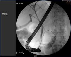
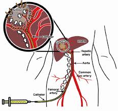
TIPS is a procedure where your doctor creates an artificial channel inside the liver to reduce liver pressure( Portal hypertension).This modality efficiently treats portal hypertension and is life saving in cases with massive gastrointestinal bleeding in those with cirrhosis liver .TIPS in nut shell shunts blood from intestines to heart.

How does the procedure work?

When you are sleeping, puncture of your jugular vein on the right side of your neck is done with a needle and then insert tube like devices into the neck vein which will ultimately reach liver. Once inside liver, a stent or stent graft will be placed between the portal and hepatic vein to create a lasting connection.
You will be conscious but sedated for the procedure, placement of the STENT which is used to shunt blood will be done using fluoroscopy and ultrasound for guidance.

Why perform it?

The TIPS procedure is usually performed in patients with liver cirrhosis. If you have this condition, your normal blood flow through the liver is blocked by scar tissue within the liver, which increases the pressure in your abdominal veins. The veins in your gullet get swollen up and once they rupture, vomiting of blood results.
Another symptom of liver cirrhosis is an abnormal collection of fluid (ascites) in the abdominal cavity.
You may be advised to undergo TIPS if you have varices which bleed acutely or recurrently and have not responded to other treatments.
You may be advised to undergo TIPS if you have varices which bleed acutely or recurrently and have not responded Once the TIPS stent is placed, the pressure in the abdominal(Portal) vein decreases, protecting the area from bleeding and reducing the water logging inside the abdomen.other treatments.

FIBROSCAN

Fibroscan is a non invasive technique to assess progression of fatty liver disease and long standing liver disease due to many other causes.It is a non invasive painless procedure, very much similar to routine ultrasonographic scanning.It is done as a day care procedure with approximate scanning time of 15-20 minutes.
Fibroscan is a specialised ultrasonologic technique which looks at the liver stiffness and gives an estimate of liver scarring. The same fibroscan machine gives a rough estimate of quantum of fat in the liver- the measurement is known as controlled attenuation parameter(CAP).
In fatty liver disease, to begin with bland fat accumulates in the liver and over time indices inflammation or fatty hepatitis. This inflammation ultimately culminates in scar tissue formation or fibrosis.
The fibrosis result is measured in kilopascals (kPa) It’s normally between 2 and 6 kPa. The highest possible result is 75 kPa. Many people with liver disease(s) have a result that’s higher than the normal range. Based on this value your liver doctor would tell you how advanced is the liver scarring process and whether you have already reached the stage of advanced scarring called liver cirrhosis.
Your CAP score is a measurement of fatty change in your liver. Your healthcare provider will use your CAP score to find out your steatosis grade.
The CAP score is measured in decibels per meter (dB/m). It ranges from 100 to 400 dB/m. CAP score categorises fatty liver into three grades- S1,S2 and S3.
CAP score
Grade of fat in liver
Approximate percentage of fat in liver
238 to 260dB/m
S1
11 to 33%
260 to 290 dB/m
S2
More than 290dB
S3
67% or more

Intragastric Balloon (IGB)

Intragastric balloon placement is a weight-loss procedure that involves placing a saline-filled silicone balloon in your stomach. This helps you lose weight by limiting how much you can eat and making you feel fuller faster.
The intragastric balloon procedure may be an option if you have concerns about your weight, and diet and exercise haven’t worked for you.
Like other weight-loss procedures, an intragastric balloon requires commitment to a healthier lifestyle. You need to make permanent healthy changes to your diet and get regular exercise to help ensure the long-term success of the procedure.

Why perform it?

The placement of an intragastric balloon helps you lose weight. Weight loss can lower your risk of potentially serious weight-related health problems, such as:
Intragastric balloon placement and other weight-loss procedures or surgeries are typically done only after you’ve tried to lose weight by improving your diet and exercise habits.
Copyright © 2019 – All rights reserved – Comprehensive Liver Care Trust – C.L.C